
Buy Now the Hard Copy of notes for seamless and ad-free learning, Click Below!
The gastrointestinal (GI) tract, also known as the digestive tract, is a continuous tube that runs from the mouth to the anus.
It is responsible for breaking down food, absorbing nutrients, and eliminating waste.

Anatomy of GI Tract Here's a brief overview of the four primary layers of the gastrointestinal tract (GIT) from the innermost to the outermost layer:
1) Mucosa:
The innermost layer responsible for secretion, absorption, and protection.
It has three sub-layers: epithelium, lamina propria, and muscularis mucosae.
2) Submucosa:
A layer of connective tissue that contains blood vessels, lymphatic vessels, nerves, and glands.
3) Muscularis externa:
A layer of smooth muscle that propels food through the GIT. It has an inner circular layer, an outer longitudinal layer, and an additional oblique layer in the stomach.
4) Serosa/Adventitia:
The outermost layer, composed of connective tissue and mesothelium (serosa) or connective tissue (adventitia) that anchors the GIT to surrounding structures.
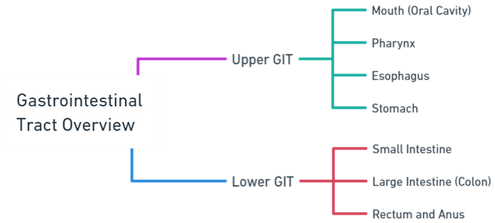
Here's a mind map overview of the Gastrointestinal Tract
Upper GI Tract
1) Mouth (Oral Cavity)
Begins the digestive process.
Structures include the lips, cheeks, palate, tongue, teeth, and salivary glands.
Salivary glands (parotid, submandibular, and sublingual) produce saliva that moistens food and begins the digestion of carbohydrates with the enzyme amylase.
2) Pharynx
A muscular tube that connects the mouth to the esophagus.
Plays a role in swallowing, directing food from the mouth to the esophagus, and preventing food from entering the trachea (windpipe) through the epiglottis.
3) Esophagus
A muscular tube about 10 inches long, running from the pharynx to the stomach.
Transports food to the stomach via peristaltic movements.
The lower esophageal sphincter (LES) controls the passage of food into the stomach and prevents the backflow of gastric contents.
4) Stomach
A J-shaped muscular organ that stores, mixes, and digests food.
Divided into four regions: cardia, fundus, body (corpus), and pylorus.
The stomach lining contains gastric glands that secrete gastric juice, including hydrochloric acid and digestive enzymes like pepsin.
Lower GI Tract
1) Small Intestine
Approximately 20 feet long, divided into three sections: duodenum, jejunum, and ileum.
The duodenum receives chyme from the stomach, along with bile from the liver and gallbladder, and pancreatic juices from the pancreas, which aid in digestion.
The jejunum and ileum are mainly involved in the absorption of nutrients and water.
2) Large Intestine (Colon)
About 5 feet long, divided into the cecum, ascending, transverse, descending, and sigmoid colon, and the rectum.
Absorbs remaining water and electrolytes from indigestible food matter and forms solid waste (feces).
The colon houses a large number of bacteria that play a role in the fermentation of undigested food, vitamin production, and protection against harmful bacteria.
3) Rectum and Anus
The rectum stores feces until they are expelled from the body.
The anus is the final part of the GI tract, consisting of internal and external sphincters that control the expulsion of feces.
Accessory Digestive Organs
In addition to the primary GI tract, several accessory organs contribute to the digestive process:
1) Salivary Glands
As mentioned, these include the parotid, submandibular, and sublingual glands, which secrete saliva to initiate the digestion of carbohydrates.
2) Liver
The body's largest gland, playing a crucial role in metabolism, detoxification, and the production of bile, which helps in the digestion and absorption of fats.
3) Gallbladder
Stores and concentrates bile from the liver, releasing it into the duodenum to aid in digestion.
4) Pancreas
Produces pancreatic juice containing digestive enzymes and bicarbonate ions, which neutralize the acidity of chyme entering the duodenum from the stomach.
Buy Now the Hard Copy of notes for seamless and ad-free learning, Click Below!

