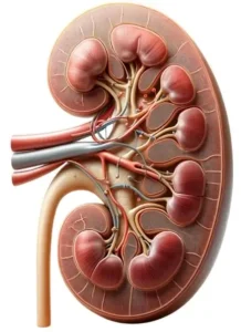
- kidney anatomy is intricate, designed to efficiently filter blood, remove waste, maintain electrolyte balance, and regulate blood pressure.
- Each kidney is a bean-shaped organ, approximately the size of a fist, located on either side of the spine, just below the rib cage.
- The kidneys are part of the upper urinary tract and play a crucial role in the body’s urinary system.

External Anatomy
-
Location
- The kidneys are retroperitoneal organs, meaning they are located behind the peritoneum (the lining of the abdominal cavity).
- They lie on either side of the vertebral column, with the right kidney positioned slightly lower than the left due to the presence of the liver.
-
Size and Shape
- Each kidney is approximately 12 cm (4.5 inches) long, 6 cm (2.5 inches) wide, and 3 cm (1.2 inches) thick, resembling a large bean.
-
Renal Capsule
- A tough, fibrous membrane surrounding each kidney, protecting it from trauma and infection.
-
Renal Hilum
- The medial indentation where the renal artery enters, and the renal vein and ureter exit the kidney. It acts as a gateway for blood vessels and nerves.
Internal Anatomy:
-
Renal Cortex
- The outer layer of the kidney, granular in appearance, containing glomeruli, proximal and distal convoluted tubules, and blood vessels.
-
Renal Medulla
- The inner layer, consisting of 10 to 18 renal pyramids. These cone-shaped tissues house the loops of Henle and collecting ducts. The apex of each pyramid, called the renal papilla, points towards the renal pelvis.
-
Renal Columns
- Inward extensions of cortical tissue separating the renal pyramids. These columns provide a path for blood vessels to enter and exit the cortex.
-
Renal Pelvis
- A funnel-shaped space in the center of the kidney that collects urine from the major calyces and channels it into the ureter.
-
Calyces
- The renal pelvis is divided into major calyces, which further divide into minor calyces. These cup-shaped structures enclose the tips of the renal pyramids, collecting urine and draining it into the renal pelvis.
Blood Supply:
-
Renal Arteries
- Branches of the abdominal aorta that deliver oxygenated, nutrient-rich blood to the kidneys.
-
Renal Veins
- Carry deoxygenated, filtered blood from the kidneys back to the inferior vena cava.
- The kidneys receive a significant amount of blood flow, essential for their filtration and excretion functions.
Microscopic Structure:
-
Nephrons
- The functional units of the kidney, with approximately 1 to 1.5 million nephrons per kidney.
- Each nephron contains a glomerulus (a capillary bed that filters blood) and a renal tubule, where the filtrate is processed and converted into urine.

