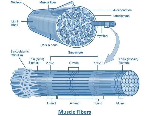
- Muscle contraction is a complex process that involves the interaction of myofilaments (actin and myosin) within muscle fibers, leading to muscle shortening and force generation.
- The process can be broken down into four key steps: neuromuscular transmission, excitation-contraction coupling, cross-bridge cycling, and muscle relaxation.
Neuromuscular Transmission
- Action Potential Initiation: Muscle contraction begins with an action potential (nerve impulse) generated in a motor neuron.
- Acetylcholine Release: The action potential travels down the motor neuron’s axon to the neuromuscular junction (NMJ), where it triggers the release of acetylcholine (ACh).
- Muscle Action Potential: ACh diffuses across the synaptic cleft and binds to receptors on the muscle fiber’s sarcolemma (plasma membrane), generating an action potential in the muscle fiber.
Excitation-Contraction Coupling
- Propagation of Action Potential: The action potential spreads along the sarcolemma and down the T-tubules, invaginations of the sarcolemma that reach deep into the muscle fiber.
- Calcium Release: This electrical signal triggers the release of calcium ions from the sarcoplasmic reticulum (SR) into the sarcoplasm (the cytoplasm of the muscle fiber).
- Calcium Binding: Calcium ions bind to troponin on the thin filaments (actin), causing a conformational change that moves tropomyosin and exposes the myosin-binding sites on actin.
Cross-Bridge Cycling
- Cross-Bridge Formation: Myosin heads (cross-bridges) on the thick filaments bind to the exposed sites on actin.
- Power Stroke: Myosin heads, energized by the hydrolysis of ATP, pivot and pull the actin filaments toward the center of the sarcomere, shortening the muscle.
- Release and Reset: Myosin heads release ADP, detach from actin when a new ATP binds, and return to their original position after ATP is hydrolyzed, ready to form another cross-bridge.
- Continuation: This cycle repeats as long as calcium ions remain bound to troponin and ATP is available.
Muscle Relaxation
- Cessation of Neural Signal: Muscle relaxation occurs when the motor neuron stops releasing ACh, halting the action potential in the muscle fiber.
- Calcium Reuptake: Calcium ions are pumped back into the SR, reducing calcium levels in the sarcoplasm.
- Blocking of Myosin-Binding Sites: As calcium dissociates from troponin, tropomyosin moves back to block the myosin-binding sites on actin, preventing further cross-bridge formation.
- Relaxation: The muscle fiber returns to its resting state.


