
Introduction to Capillary Electrophoresis (CE)
- Capillary Electrophoresis (CE) is a sophisticated electrophoretic technique that utilizes narrow capillaries to achieve high-resolution separations with rapid analysis times.
- CE is highly automated and suitable for a wide range of applications, including the analysis of small ions, biomolecules, and pharmaceuticals.
Principle of Capillary Electrophoresis (CE)
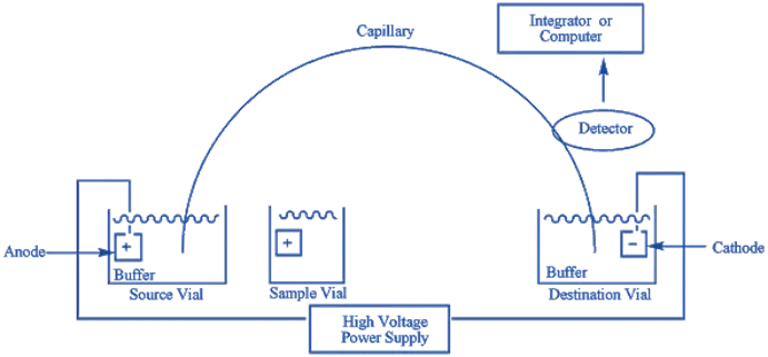
- In CE, an electric field is applied across a thin capillary filled with an electrolyte, causing charged molecules to migrate based on their electrophoretic mobility.
- The small diameter of the capillary reduces band broadening, enhancing separation efficiency.
Advertisements
Methodology
- Capillary Preparation: A fused silica capillary is cleaned and conditioned before use.
- Sample Injection: Small volumes of the sample are introduced into the capillary via electrokinetic or pressure-based methods.
- Buffer Filling: The capillary is filled with an appropriate running buffer.
- Application of Electric Field: A high-voltage power supply drives the migration of analytes through the capillary.
- Detection: As molecules exit the capillary, they are detected using methods like UV absorbance, fluorescence, or mass spectrometry.
- Data Analysis: The resulting electropherogram is analyzed to determine the presence, concentration, and characteristics of the analytes.
Modes of Capillary Electrophoresis (CE)
- Capillary Zone Electrophoresis (CZE): Separates analytes based on charge-to-size ratio in a buffer-filled capillary.
- Capillary Gel Electrophoresis (CGE): Uses a gel medium for separating DNA fragments.
- Micellar Electrokinetic Chromatography (MEKC): Employs micelles to separate neutral and charged analytes.
- Capillary Isoelectric Focusing (cIEF): Separates proteins by their isoelectric point (pI).
- Capillary Electrochromatography (CEC): Combines CE and HPLC, using chromatographic material for separation.
Detection Methods
- UV/Vis Absorption: Common, requires UV-active analytes.
- Fluorescence: Detects native or tagged fluorescent compounds.
- Mass Spectrometry (MS): Coupled with CE for precise molecular identification.
Advertisements
Advantages
- High Resolution: Exceptional separation efficiency due to minimal band broadening.
- Speed: Rapid analysis times, often completed within minutes.
- Small Sample Volume: Requires only nanoliter to microliter sample volumes.
- Automation: Highly automated systems allow for high-throughput analyses.
- Versatility: Can analyze a broad range of analytes, including ions, small molecules, and biomolecules.
Disadvantages
- Complexity: Requires specialized equipment and expertise.
- Cost: High initial investment for instrumentation and maintenance.
- Sensitivity: May require sensitive detection methods for low-abundance analytes.
- Buffer Requirements: Strict buffer conditions are necessary to maintain capillary integrity and performance.
- Sample Preparation: Some samples may require extensive preparation to be compatible with CE.
Applications
- Pharmaceutical Analysis: Drug purity testing and pharmacokinetic studies.
- Clinical Diagnostics: Analyzing biomarkers and metabolic profiles.
- Genomics and Proteomics: High-throughput DNA and protein analysis.
- Environmental Monitoring: Detection of pollutants and toxins.
- Forensic Science: Identification and quantification of substances in forensic samples.
Advertisements